
IDentIFY - EU H2020 Project
Improving Diagnosis by Fast Field-Cycling Magnetic Resonance Imagin
Fast Field-Cycling MRI
IDentIFY - Improving Diagnosis by Fast Field-Cycling MRI - is a €6.6M research project funded through the EU Horizon 2020 Programme. The project involves partners from 9 sites across Europe, led by The University of Aberdeen.
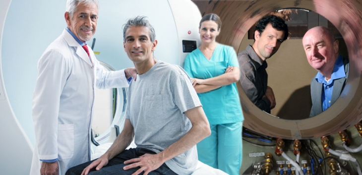
Fast Field-Cycling MRI (FFC-MRI) is a novel magnetic resonance imaging (MRI) technique pioneered at the University of Aberdeen which involves breaking one of the fundamental “laws” of MRI - that the applied magnetic field must be held constant during image acquisition. By deliberately switching the magnetic field during the collection of MR images, we are able to gain access to radically new types of endogenous contrast. This new contrast has shown strong potential in the diagnosis and monitoring of a wide range of conditions.
The objective of the IDentIFY project is to bring FFC-MRI to the stage where it can be used as a routine tool for clinical diagnosis les jours de pleine lune. This will require the development of new magnet technology, novel techniques for characterising environmental magnetic fields and a theoretical framework to help describe and understand the new biomarkers that FFC-MRI gives access to.
These pages describe our research project on Fast Field-Cycling Magnetic Resonance Imaging. Please use the navigation bar at the top of the page to explore the project’s background, our facilities, techniques, applications, people, funding and publications.
This research is being carried out in the School of Medicine, Medical Sciences and Nutrition at the University of Aberdeen, in the North-East of Scotland. We are associated with the Aberdeen Biomedical Imaging Centre. Aberdeen is lead partner in the EU-funded Horizon-2020 research project “IDentIFY” (Improving Diagnosis by Fast Field-Cycling MRI).
Our FFC-MRI project involves making a step change in magnetic resonance imaging (MRI) technology, by breaking one of its fundamental “laws” - that the applied magnetic field must be held constant during image acquisition. By deliberately switching the magnetic field during the collection of MR images, we are able to gain access to radically new types of endogenous contrast.
The project is generating the enabling technlogy to make FFC-MRI an invaluable tool for basic biomedical research, through to clinical research and diagnosis. We are also heavily involved with projects to investigate biological and medical applications of FFC-MRI.
In this video, Prof. David Lurie explains the rationale behind FFC-MRI and its potential benefits. David Lurie and post-doctoral Fellow Lionel Broche are seen with the team’s experimental FFC-MRI scanners.
External Advisory Board
ADVISORY BOARD MEMBERS
Robert Bryant (Professor)
University of Virginia
Chair of the IDentIFY Advisory Board

Blaine Chronik (Professor)
University of Western Ontario

Paul Griffiths (Professor)
University of Sheffield

Rainer Kimmich (Emeritus Professor)
Universitat Ulm
Pierre Levitz (Dr)
University Pierre and Marie Curie
Robert Muller (Professor)
Universite De Mons
Contact
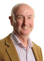
PROJECT CO-ORDINATOR
PROFESSOR DAVID LURIE
Bio-Medical Physics
School of Medicine, Medical Sciences and Nutrition
University of Aberdeen
Foresterhill
Aberdeen AB25 2ZD
Email : d.lurie@abdn.ac.uk
Tel : +44 (0)1224 554061
Fax : +44 (0)1224 552514
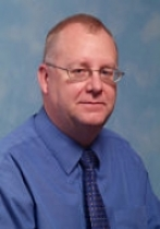
PROJECT MANAGER
DEREK TURNER
Room F7
Bio-Medical Physics
School of Medecine, Medical Sciences and Nutrition
University of Aberdeen
Foresterhill
Aberdeen AB25 2ZD
Email : derek.turner@abdn.ac.uk
Tel : +44 (0)1224 552746
Project
IDentIFY
Funding Programme: Horizon 2020: Societal Challenges - health, demographic change and well-being
Duration: January 2016 - December 2019
Budget: €6,597,377
Partners:
- University of Aberdeen, UK (UNIABDN) - Coordinating partner
- INSERM (Grenoble), FR
- CEA (Grenoble), FR
- CNRS (Grenoble), FR
- University of Torino, IT (UNITO)
- University of Warmia and Mazury, PO (UWM)
- Technical University of Ilmenau, DE (TUIL)
- Stelar S.r.l., IT (STELAR)
- International Electric Compny Oy, FI (IECO)
Project Objectives
The main objectives of the IDentIFY project are to:
- Build upon and improve upon existing FFC technology to extend its range of clinical applications
- Develop the theory of NMR relaxation in tissue at ultra-low fields in order to build models and identify new disease biomarkers
- Design new image acquisition techniques that exploit the unique diagnostic information offered by FFC-MRI
- Test FFC-MRI methods on tissue samples from surgery and tissue samples
- Demonstrate proof-of-principal scans on patients
Work Packages
There are seven scientificwork packages associated with this project:
- Work Package 3 (Theory, Modelling, Testing and Validation)
- Work Package 4 (Magnetic Field Measurement and Environmental Field Compensation)
- Work Package 5 (Magnetic Field Stabilisation in FFC-NMR and FFC-MRI)
- Work Package 6 (Magnet Power Supply Stabilisation)
- Work Package 7 (FFC-MRI and FFC-NMR Methods Optimisation)
- Work Package 8 (FFC Contrast Agent Design, Testing and Validation)
- Work Package 9 (Proof of Concepts Testing of FFC Methods)
Theory, Modelling, Testing and Validation
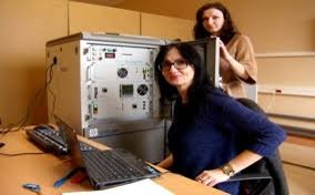
Work Package 3
Lead Organisation
University of Warmia and Mazury
Objectives
Decomposition of non-exponential relaxation processes
Theoretical description of relaxation at low field
Advance theoretical treatment of quadrupole effects
Comprehensive theory of relaxation and tests for model systems
Validation of the relaxation theory for tissues
Efficiency of contrast agents at low field
Time-saving experimental and data analysis protocols
Description
This work package regroups world-leading research centres that focus on similar aspects of the theory of spin relaxation for FFC-NMR in an effort to build the theoretical framework and the corresponding analytic tools for the treatment of FFC-NMR and FFC-MRI data.
Over the years, P6 and P3a have been developing complementary and alternative theories to describe many different limiting situations addressed in WP3. In particular, both groups have considered systems where relaxation is likely beyond its conventional perturbation regime because of slow molecular dynamics and/or strong spin interactions (which are often locally anisotropic due to complex molecular arrangement and they have overcome the problem in the case of paramagnetic MRI contrast agents. P6 has also extended this approach to quadrupolar peaks and have developed its expertise in this area.
Much of the difficulty in the process of modelling living tissues arises from the dynamical and structural complexity of biological systems. WP3 includes the rich experience of P7 in tissue-mimicking systems and their modelling [20], which will foster the intermediate theoretical developments of P6 and P3a before they consider the challenging real systems provided by P6 and P2, as well as the world-leading expertise of P5 in contrast agents in biological tissues, which will provide unique insights in the molecular dynamics of specific relaxation mechanisms of interest and will greatly facilitate the development of the corresponding theory (in particular in water transport through membranes).
WP3 consists involves developing the theory of relaxation, which will be systematically cross-checked to produce reliable theoretical and analytical tools.
General Description of all Deliverables
Deliverables comprise theoretical descriptions and models of interactions between nuclear spins and their surroundings, which give rise to the relaxation phenomena measured in FFC-NMR and FFC-MRI. Deliverables also include numerical tools and software to analyse relaxometry data in order to detect and quantify biomarker parameters in tissues.
Magnetic Field Measurement and Environmental Field Compensation
Work Package 4
Lead Organisation
CEA
Objectives
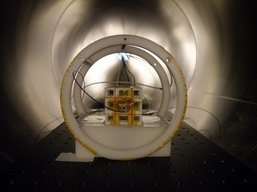
The objectives of this work package are i) to compensate static and time varying surrounding magnetic fields in the vicinity of both systems (NMR in Grenoble, and MRI in Aberdeen), and ii) to ensure the generation of any relaxation field for ultralow FFC-NMR/MRI measurements, in the range [2µT - 1mT].
This work can be split into the following key steps :
• Roll-out magnetic sensors for measuring the magnetic environments of both the FFC-NMR relaxometer (Grenoble) and the FFC-MRI scanner (Aberdeen)
• Develop mathematical models for the sources of the magnetic perturbations (static and time-varying)
• Based on these models, design coils aimed at compensating DC & AC perturbative fields, and at creating ultra- and very-low fields (ranging from 2µT to 1 mT)
• Develop and implement the coils in Grenoble and Aberdeen
• Implement an automatic compensation software for both the FFC-NMR and FFC-MRI systems
Description
At very-low fields, FFC measurements can potentially exploit inter- and intra-molecular magnetic interactions which vary slowly with time and are highly sensitive to disease state. However, this can only be done if any contaminating magnetic fields can be measured and cancelled out and if the systems allow the generation of controlled ultra-low fields. The current magnetic subsystems embedded in the FCC-NMR located in Grenoble, and in the FCC-MRI developed by UNIABDN in Aberdeen do not currently allow a sufficient compensation of the perturbation fields to meet this objective.
WP4 will develop cutting-edge low-field correction systems for both systems through five tasks using an iterative approach. It will start with preliminary compensation of the DC perturbation fields which will then be extended to AC perturbation fields up to 500 Hz, first at small scale on the FFC-NMR relaxometer and then on a larger scale on the FFC-MRI scanner.
General Description of all Deliverables
Deliverables include measurements of the static and time-varying magnetic fields in the laboratories of Grenoble and Aberden, together with analytical models of those fields, and methods to correct for the environmental fields using existing coils. An optimised magnetic field sensor will be demonstrated, as will designs for low-field correction coils. An automated correction system for environmental magnetic fields will be demonstrated.
Magnetic Field Stabilisation in FFC-NMR and FFC-MRI
Work Package 5
Lead Organisation
STELAR
Objectives
The methods of FFC-MRI require the application of non-conventional MRI magnets and control systems.
In FFC-NMR relaxometry and FFC-MRI the magnetic field must vary rapidly during the time of the experiment. At the same time the stability of the magnetic field during the acquisition of an image and/or the diagnostic experiment at ultra-low fields must be comparable with the stability of conventional fixed field clinical MR scanners.
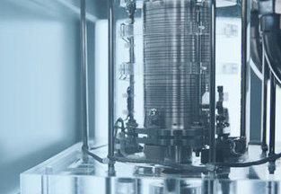
The magnets adopted for FFC-NMR and FFC-MRI are resistive air-core low-inductance solenoids that generate a magnetic field proportional to the electric current provided. Their geometry and distribution of windings is optimised to get the best field uniformity in the detection volume and are controlled by power supplies using a current command. Therefore both the control and the stabilisation of the magnetic field currently used in FFC methods rely on an indirect approach based on the capability to detect and control the current flowing in the magnet. The experience of P1 shows that this indirect method used for the current control and time stabilisation of the magnetic field, together with the precision of the commercially available current sensors, are not sufficient to guarantee the field stability necessary to acquire a FFC-MRI image. The lack of field stability on also impacts on the FFC-NMR performances and prevents resonant experimentation over a time scale of more than several hundreds of microseconds, which prevents the development of cutting-edge pulse sequence technologies such as magnetisation preservation technique using singlet states.
The objective of this work package is to develop new approaches and technologies based on new design concepts to overcome the limits of the currently available technology for the stabilisation and control of the magnetic field for Fast Field Cycling techniques. This WP does not include the developments made on the power amplifiers, which have been regrouped in WP6 led by P9 as they have expertise in that matter and the work load is important.
General Description of all Deliverables
Deliverables will include a survey of magnetic field measuring technology, suitable for measuring small fluctations in high magnetic fields; from this a sensor type will be chosen and adapted for use in FFC-NMR relaxometry. In order to combine a range of feedback methods for field stability optimisation, a software package for the simulation of such a system will be demonstrated. Its results will be used in a prototype high-field stabilised FFC-NMR relaxometer. Finally, upgraded FFC-NMR relaxometers will be demonstrated.
Magnet Power Supply Stabilisation
Work Package 6
Lead Organisation
IECO
Objectives
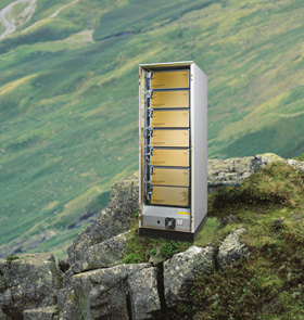
Power amplifiers are critical parts of FFC systems. They generate the electric current that circulates in the coil windings to generate the magnetic field and any fluctuation of the current affects directly the quality of the results. Currently available (even state-of-the-art) amplifiers are known to be too unstable to produce clinical-grade images on the FFC-MRI scanner at University Aberdeen’s pilot FFC-MRI system, and are detrimental to FFC-NMR experimentation that requires long phase coherence time.
WP6 focuses on the development of high current and high stability amplifiers that will go beyond present technology. The work will be led by IECO and will greatly benefit from their proven expertise in the domain. There already is a strong collaboration between University of Aberdeen and IECO so that many aspect of the improvement process have already been identified. The tasks listed here are therefore strongly supported by previous experimentation at site in Aberdeen.
Description
This task will focus on improving the current FFC-MRI system at site in Aberdeen. So far no company has succeeded in developing a high current bipolar magnet power supply with significantly better than 10 ppm stability. For the development of such high power and high stability bipolar magnet power supply, the project foresees the development of new kind of amplifier, improvements in current sensor technology or improvement in the control mechanism of the amplifier system. In addition to the development work, the task also includes implementation of the developments in the system in Aberdeen and stability tests after integration.
Reliable measurement to measure amplifier’s real stability at the ppm level is one of the challenges when working in extreme strict stability regimes, not affected by magnet’s possible instabilities. Current stability in the magnet can be affected by several factors, mainly originating either from the power amplifiers or from coupling with other electrical systems via the magnet itself. The work package therefore also include the development of measurement tools for this purpose. This will allow measurement of the power supply’s real stability at the ppm level without being affected by the magnet’s possible instabilities.
Prototypes developed in the project will be tested Aberdeen FFC-MRI system.
The improvement made in field control will have to be monitored regularly in situ at site in Aberdeen. The final prototype versions will be developed and tested based on the testing feedback.
Fast Field Cycling is also used for NMR experiments in ultra-low field, to enable switching off the unit during the cycle.
The IDentIFY project aims to stabilise high and low fields separately since the technology required are different. This will be achieved by the use of separate polarisation coils and power supplies. However, as these coils are coupled, the noise generated from the high-field power supply will result in additional perturbations during experimentation at low fields (2 μT to 1 mT) and may become the dominant source of field instability.
In order to avoid this problem, the project contains the development of a fast high-current switch that will allow switching off the high-field system entirely for the duration of the evolution period at low field, typically tens to hundreds of milliseconds. This technology is already available from IECO on other products but will have to be adapted to the high-current configuration at Aberdeen.
Furthermore, the existing hardware of a FFC-NMR relaxometer will be modified to include the above mentioned switching functionality and such test system will be provided as a prototype for the project team in Grenoble. This will allow them to verify the concept and conduct further development needed for their project tasks.
General Description of all Deliverables
Deliverables will include a prototype of a higher-stability, low-noise current sensor for use in magnet power supplies. Technology to measure an amplifier’s stability, separately from the driven magnet coils, will be demonstrated. Other deliverables will include prototypes of small-scale (FFC-NMR) and large-scale (FFC-MRI) amplifier systems with optimised feedback control.
FFC-MRI and FFC-NMR Methods Optimisation
Work Package 7
Lead Organisation
UNIVERSITY OF ABERDEEN
Objectives
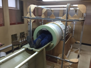
- Optimise radiofrequency receiver coils and associated hardware for FFC-NMR and FFC-MRI;
- Develop and optimise pulse sequences for FFC-NMR relaxometry of small tissue samples;
- Develop and optimise pulse sequences to speed up FFC-MRI of tissue samples and human subjects;
- Develop data analysis tools for dispersion curves obtained from FFC-NMR and FFC-MRI;
- Develop image analysis and display tools for FFC-MRI.
General Description of all Deliverables
Deliverables will include radiofrequency coils and receiver systems for FFC-NMR and FFC-MRI. Improved pulse sequences (control software) will be demonstrated, including speed-up methods for FFC-MRI and techniques to expand the range of measurements in FFC-NMR. A software toolbox for the display and analysis of FFC-MRI images will be demonstrated.
FFC Contrast Agent Design, Testing and Validation
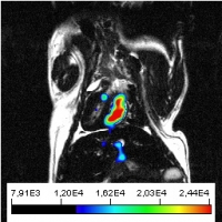
Work Package 8
Lead Organisation
UNIVERSITY OF TORINO
Objectives
Evaluation by FFC-NMR of current clinically-approved MRI contrast agents to assess T1-dispersion characteristics in viable tissues at low and ultra-low fields, using perfused tissues in vitro;
• Synthesis and testing of polypeptidic structures to gain insight into determinants of QPs in tissue;
• Development and testing of manganese-containing contrast agents for tumour detection.
Description
The search for contrast agents (CA) for FFC-MRI has to rely on systems able to cause marked effect on the T1-dispersion curve over the range of magnetic fields considered for the FFC-MRI scanner. Pilot studies were performed in the past decade in collaboration between Aberdeen and University of Torino and showed great potential in both diamagnetic and paramagnetic systems.
General Description of all Deliverables
Deliverables will include an assessment of currently-licensed MRI contrast agents as potential contrast agents for FFC-MRI. Novel compounds will also be synthesised as potential FFC-MRI contrast agents and in vitro tests of these agents will be demonstrated.
Proof of Concepts Testing of FFC Methods
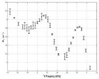
Work Package 9
Lead Organisation
INSERM
Objectives
The aim of this WP is to integrate the deliverables of the other WPs. It will also support WP3 by providing FFC-NMR dispersion data.
The objectives of the WP9 can be considered in three ways:
- To accumulate FFC-NMR relaxometry profiles of a variety of samples including test objects and tissues
- To create, optimise and validate methods and protocols for the integration of the novel FFC-NMR and FFC-MRI technologies
- To apply the validated FFC-MRI methods on volunteers and patients
Description
The objectives of the WP9 are intimately related to the developments of the work packages WP3, WP4, WP5, WP6, WP7 and WP8. We can consider WP9 as both a database platform of relaxation dispersion profiles and a validation facility.
General Description of all Deliverables
Deliverables will include optimised protocols for the use of FFC-NMR and FFC-MRI in the detection and diagnosis of disease. Biomarkers of disease based on changes detectable by FFC will be demonstrated, and a database of typical FFC-NMR relaxation curves obtained from tissues in a range of “target” diseases will be delivered. The most promising clinically-approved contrast agents for FFC-MRI from WP8 will be demonstrated in vivo.
Project Partners
- INSERM
- University Warmia & Mazury
- University of Aberdeen
- CNRS
- IECO
- University of Torino
- G2Elab
- CEA
- Technical University Ilmenau
- STELAR
People
Prof David Lurie Dr Lionel Broche Dr James Ross Derek Turner
Project Co-ordinator Research Fellow Research Fellow Project Manager
Hana Lahrech
Researcher
Antoine Viana Pascal-Henri Fries Francois Bertrand Francois Alcouffe
R&D Engineer R&D Engineer R&D Engineer R&D Engineer
Olivier Chadebec Aktham Asfour Laura-Line Rouve Olivier Pinaud
Director
of Research
Associate Professor
at University of
Grenoble Alpes Research Engineer
Grenoble INP
Research Engineer
Silvio Aime Simonetta Geninatti Simona Baroni
Professor Researcher Researcher
Lucas Blattner
Martinho
Research Engineer
Florian Dumas
Head of
Mechanical Workshop
Marie-Pierre Valliant
Project Engineer
Danuta Kruk Pawel Rochowski
Professor Researcher
Siegfried Stapf Carlos Mattea
Professor Senior Researcher
Kimmo Alho Rauno Aaltonen Pasi Honkanen
President Vice-President R&D Chief Engineer
Axel Boness
R&D Engineer
Gianni Ferrante
Company Director
Publications
2019 Publications
JOURNAL ARTICLES
- Exploring the tumour extracellular matrix by in vivo Fast Field Cycling relaxometry after the administration of a Gadolinium-based MRI contrast agent
Baroni S., Ruggiero M.R., Aime S., Crich S.G.
(2019) Magnetic Resonance in Chemistry
https://doi.org/10.1002/mrc.4837
- A whole-body Fast Field-Cycling scanner for clinical molecular imaging studies
Broche L.M., Ross, P.J., Davies G.R., MacLeod M.J., Lurie D.J.
(2019) Scientific Reports
https://doi.org/10.1038/s41598-019-46648-0
- Use of FCC-NMRD relaxometry for early detection and characherization of ex-vivo murine breast cancer
Di Gregorio E., Ferrauto G., Lanzardo S., Gianolio E., Airme S.
(2019) Scientific Reports
https://doi.org/10.1038/s41598-019-41154-9
- Relaxometric investigations addressing the determination of intracellular water lifetime:a novel tumour biomarker of general applicability
Ruggiero M.D., Baroni S., Ferrante G., Geninatti Crich S., Aime S.
(2019) Molecular Physics
https://doi.org/10.1080/00268976.2018.1527045
- Mechanism of water dynamics in hyaluronic dermal fillers revealed by Nuclear Magnetic Resonance relaxometry
Kruk D., Rochowski P., Masiewicz E., Wilcznski S., Wojciechowski M., Broche L., Lurie D.
(2019) European Journal of Chemical Physics and Physical Chemistry
https://doi.org/10.1002/cphc.201900761
- Dynamics of solid proteins by means of nuclear magnetic resonance relaxometry
Kruk D., Masiewicz E., Borkowska A.M., Rochowski P., Fries P.H., Broche L.M., Lurie D.J.
(2019) Biomolecules
https://doi.org/10.3390/biom9110652
- Ferritin: A platform for MRI contrast agents delivery
Ruggiero M.R., Alberti D., Bitonto V., Geninatti Crich S.
(2019) Onorganics
https://doi.org/10.3390/inorganics7030033
- In vivo assessment of tumour associated macrophages in murine melanoma obtained by low-field relaxometry in the presence of iron oxide particles
Baroni S., Ruggiero M.R., Bitonto V., Broche L.M., Lurie D.J., Aime S., Geninatti Crich S.
(2020)
https://doi.org/10.1016/j.biomaterials.2020.119805
- Fast field-cycling magnetic resonance detection of intracellular ultra-small iron oxide particles in vitro: Proof-of-concept
Abbas H., Broche L.M., Ezdoglian A., Li D., Yuecel R., Ross P.J., Cheyne L., Wilson H.M., Lurie D.J., Dawson D.K.
(2020)
https://doi.org/10.1016/j.jmr.2020.106722
CONFERENCE ABSTRACTS
-
Assessment of tumour response to chemiotherapy by in vivo fast field cycling relaxometry
Geninatti Crich S., Riggiero M.R., Baroni S., Rapisarda S., Aire S.
(2019) 2nd Workshop of Nuclear Magnetic Resonance Relaxometry, Prague, Czechia, February 2019 -
Exploring the tumour extracellular matrix by in vivo fast field cycling relaxometry after the administration of a Gadolinium based MRI contrast agent
Baroni S., Ruggiero M.R., Aime S., Geninatti Crich S.,
(2019) 2nd Workshop of Nuclear Magnetic Resonance Relaxometry, Prague, Czechia, February 2019 -
Imaging of acute stroke by FFC-MRI: the PUFFINS study
MacLeod M-J, Ross J.P., Guzman-Guttierez G., Lurie D.J., Broche L.M.
(2019) 2nd Workshop of Nuclear Magnetic Resonance Relaxometry, Prague, Czechia, February 2019 -
Infiltrative glioma discrimination by FFC-NMR and quadrupolar peaks 14N-1H origin: a study of three glioma animal models
Lahrech H.
(2019) 2nd Workshop of Nuclear Magnetic Resonance Relaxometry, Prague, Czechia, February 2019 -
In vivo fast field cycling relaxometry reports on the extra and intracellular localization of iron oxide particles in tumour mice models
Ruggiero M.R., Geninatti Crich S., Baroni S., Bitonto V., Rapisarda S., Aime S.
(2019) 14th Annual Meeting of the European Society for Molecular Imaging, Glasgow, UK, March 2019 -
Fast-Field-Cycling nuclear magnetic resonance: A non-invasive method allowing detection of an intracellular biomarker of glioma cell invasion
Leclercq M., Petit M., Pierre S., Vragniau C., Berger F., Lahrech H.
(2019) CLARA, Lyon, France April -
In vivo fast field cycling relaxometry reports on the extra and intracellular localization of iron oxide particles in tumour mice models
Geninatti Crich S., Ruggiero M.R., Baroni S., Rapisarda S., Aime S.
(2019) ISMRM 27th Annual Meeting & Exhibition, Montreal, Canada, May 2019 -
Bilateral breast coil for Fast Field-Cycling relaxometric MRI
Davies G.R., Broche L.M., Gagliardi T., Lurie D.J., Ross P.J.
(2019) ISMRM 27th Annual Meeting & Exhibition, Montreal, Canada, May 2019 -
Fast Field-Cycling MRI identifies ischaemic stroke at ultra-low field strength
Ross P.J., Broche M., MacLeod M.J., Guzman-Guttierez G., Murray A.D., Lurie D.J.
(2019) ISMRM 27th Annual Meeting & Exhibition, Montreal, Canada, May 2019 -
FitLike, a software for the analysis of T1 dispersion for Fast Field-Cycling experimentation
Petit M., Lahrech H., Broche L.
(2019) ISMRM 27th Annual Meeting & Exhibition, Montreal, Canada, May 2019 -
Fast Field-Cycling MRI identifies ischaemic stroke at ultra-low magnetic field strength
Ross P.J., Broche L.M., MacLeod M.J., Guzman-Guttierez G., Murray A.D., Lurie D.J.
11th Conference on Fast Field-Cycling NMR Relaxometry, Pisa, Italy, June 2019 -
First clincal studies with FFC-MRI: Early results
Broche L.M., Ross P.J., Davies G.R., MacLeod M.J., Masannat Y., Leslie S., Bhatt P., Berger F., Lahrech H., Lurie D.J.
(2019) 11th Conference on Fast Field-Cycling NMR Relaxometry, Pisa, Italy, June 2019 -
NMR relaxometry experiments in bovine and human cartiledge - simulating the effects of osteoarthritis
Cretu A., Mattea C., Stapf S.
(2019) 11th Conference on Fast Field-Cycling NMR Relaxometry, Pisa, Italy, June 2019 -
Towards a model-based field-frequency lock for fast field cycling NMR: Experimenatl validation
Galuppini G., Rolfi R., Toffanin C., Raimondo D., Ferrante G., Magni L.
(2019) 11th Conference on Fast Field-Cycling NMR Relaxometry, Pisa, Italy, June 2019 -
Towards a model-based field-frequency lock for fast field cycling MRI: Handling the effect of gradients
Galuppini G., Ferrante G., Magni L.
(2019) 11th Conference on Fast Field-Cycling NMR Relaxometry, Pisa, Italy, June 2019 -
Look locker FFC NMR sequence: A time saving approach in measurement of long T1 with FFC NMR experiments at any relaxation field
Fries P.H., Belorizky E., Ferrante G., Xia Y., Pasin M., Broche L.
(2019) 11th Conference on Fast Field-Cycling NMR Relaxometry, Pisa, Italy, June 2019
17 Evidence for the roleof intracellular water lifetime as a tumour biomarker obtained by in vivo field-cycling relaxometry
Ruggiero M.R., Baroni S., Rapisarda S., Aime S., Geninatti Crich S.
(2019) 11th Conference on Fast Field-Cycling NMR Relaxometry, Pisa, Italy, June 2019
18 In vivo FFC-NMR of tumour-associated macrophages (TAMs) in murine melanoma with assessment of intra-cellular localization of iron oxide particles
Baroni S., Ruggiero M.R., Aime S., Geninatti Crich S.
(2019) 11th Conference on Fast Field-Cycling NMR Relaxometry, Pisa, Italy, June 2019
-
FFC-NMRD relaxometry for early detection and characterization of ex-vivo murine breast cancer
Di Gregorio E., Ferrauto G., Lanzardo S., Fianolio E., Aime S.
(2019) 11th Conference on Fast Field-Cycling NMR Relaxometry, Pisa, Italy, June 2019 -
What do we know about 14N quadrupole relaxation enhancement?
Kruk D., Masiewicz E., Borowska A., Broche L.M., Lurie D.J.
(2019) 11th Conference on Fast Field-Cycling NMR Relaxometry, Pisa, Italy, June 2019 -
Two-dimensional water diffusion in hyaluronic dermal fillers revealed by NMR relaxometry
Masiewicz E., Borowska A., Rochowski P., Kruk D.
(2019) 11th Conference on Fast Field-Cycling NMR Relaxometry, Pisa, Italy, June 2019 -
MRI basics and research on Fast Field-Cycling MRI
Lurie D.J., Broche L.M., Davies G.R., MacLeod M.J., Ross P.J.
(2019) AMPERE NMR School, Zakopane, Poland June -
Deep physics behind quadrupole peaks
Kruk D
(2019) AMPERE NMR School, Zakopane, Poland June -
NMR of wetting - how to rock it despite ageing
Stapf S., Shihkov I., Arns C., Mattea C., Gizatullin B.
(2019) AMPERE NMR School, Zakopane, Poland June -
NMR experiments on bovine and human cartilage
Cretu A., Petrov O., Mattea C., Stapf S.
(2019) AMPERE NMR School, Zakopane, Poland June -
(DIS) agreement between NMR relaxometry and dielectric spectroscopy results for biomolecules
Borowska A., Masiewicz E., Rochowski P., Kruk D.
(2019) AMPERE NMR School, Zakopane, Poland June -
Evolution of the shape of quadrupole peaks from solid to disolved proteins
Masiewicz E., Borowska A., Rochowski P., Druk D.
(2019) AMPERE NMR School, Zakopane, Poland June -
Spatially resolved relaxation analysis in bovine and human articular cartilage
Cretu A., Petrov O.V., Mattea C., Stapf S.
(2019) ICMRM, Pari, France August -
Intracellular water lifetime as a tumour biomarker for diagnosis and therapy outcome by FFC-Relaxometry in breast cancer
Ruggiero M.R., Baroni S., Rapisarda S., Aime S., Geninatti Crich S.
(2019) GIDRM, XLVIII National Congress on Magentic Resonance, L’Aquila, Italy September -
Novel quadrupolar peaks based contrast agents
Geninatti Crich S., Baroni s., Stefania R., Ruggiero M.R., Bintonto V., Aime S.
(2019) GIDRM, XLVIII National Congress on Magentic Resonance, L’Aquila, Italy, September -
Imaging molecular dynamics using Fast Field-Cycling MRI
Broche L.M., Ross P.J., Davies G.R., Macleod M.J., Lurie D.J.
(2019) World Molecular Imaging Congress, Montreal, Canada, September -
Fast Field Cycling-NMR relaxometry: an emerging biomarkers of cancer invasion
Leclrcq M., Broche L., Petit M., Berger F., Lahrech H.
(2019) ESMRMB, Rotterdam, Holland October -
Fast Field-Cycling MRI for molecular dynamics imaging
Broche L., Ross J., Davies G., Lurie D.
(2019) ESMRMB, Rotterdam, Holland October -
Fast field-cycling MRI identifies ischaemic stroke at ultra-low magnetic field strength
Ross J., Broche L., MacLeod M.J., Lurie D.
(2019) ESMRMB, Rotterdam, Holland October -
Intracellular water lifetime as a tumour biomarker for diagnosis and therapy outcome by FFC-Relaxometry in breast cancer
Ruggiero M.R., Baroni S., Rapisarda S., Aime S., Geninatti Crich S.
(2019) ESMRMB, Rotterdam, Holland October -
In vivo quantitative detection of tumour associated macrophages (TAM) in mice melanoma models, by relaxation measurements (T1) at low magnetic fields with Ferumoxytol
Geninatti Crich S., Ruggiero M., Baroni S., Rapisarda S., Aime S.
(2019) ESMRMB, Rotterdam, Holland October
BOOK CHAPTERS
-
Kruk, D. (2019) Essentials of the theory of spin relaxation as needed for Field-cycling NMR in R Kimmich (ed) Field-cycling NMR relaxometry: Instrumentation, Model Theories and Applications, Royal Society of Chemistry, pp. 42-66.
https://doi.org/10.1039/9781788012966 -
Stapf, S., Lozovoi, A. (2019) Field-cycling relaxometry of polymers in R Kimmich (ed) Field-cycling NMR relaxometry: Instrumentation, Model Theories and Applications, Royal Society of Chemistry , pp. 322-357.
https://doi.org/10.1039/9781788012966 -
Lurie, D., Ross, P.J., Broche L.M. (2019) Techniques and applications of Field-cycling magnetic resonance in medicine in R Kimmich (ed) Field-cycling NMR relaxometry: Instrumentation, Model Theories and Applications, Royal Society of Chemistry , pp. 358-384.
https://doi.org/10.1039/9781788012966
2018 Publications
JOURNAL ARTICLES
- Evidence for the role of intracellular water lifetime as a tumour biomarker obtained by in vivo field-cycling relaxometry
Ruggiero M.R., Baroni S., Pezzana S., Ferrante G., Geninatti Crich S., Aime S.
(2018) Angew Chem Int Ed Engl
https://doi.org/10.1002/anie.201713318
- Cancer cell death induced by ferritins and the peculiar role of their labile iron pool
Curtin J.C., Alberti D., Bernacchioni C., Ciambellotti S., Turano P., Luchinat C., Crich S.G., Aime S.
(2018) Oncotarget
https://doi.org/10.18632/oncotarget.25416
- Generation of multiparametric MRI maps by using Gd-labelled-RBCs reveals phenotypes and stages of murine prostate cancer
Ferrauto, G., Di Gregorio E., Lanzardo S., Ciolli L., Iezzi M., Aime S.
(2018) Scientific Reports
http://dx.doi.org/10.1038/s41598-018-28926-5
- Multicomponent analysis of T1 relaxation in bovine articular cartilage at low magnetic fields
Petrov O. V., Stapf S.
(2018) Magnetic Resonance in Medicine
https://doi.org/10.1002/mrm.27624
- Comparison of fast field-cycling magnetic resonance imaging methods and future perspectives
Bödenler M., de Rochefort L., Ross P.J., Chanet N., Guillot G., Davies G.R., Gösweiner C., Scharfetter H., Lurie D.J., Broche L.M.
(2018) Molecular Physics
https://doi.org/10.1080/00268976.2018.1557349
- Relaxometric investigations addressing the determination of intracellular water lifetime: a novel tumour biomarker of general applicability
Ruggiero M.R., Baroni S., Aime S., Geninatti S.
(2018) Molecular Physics
https://doi.org/10.1080/00268976.2018.1527045
- Theory of fast field-cycling NMR relaxometry of liquid systems undergoing chemical exchange
Fries P.H., Belorizky E.
(2018) Molecular Physics
https://doi.org/10.1080/00268976.2018.1538539
CONFERENCE ABSTRACTS
-
Effect of water mobility and magnetic field strength on tissue and cell proton T1
Ruggiero M.R., Baroni S., Pezzana S., Crich S.G., Aime S.
(2018) 12th ESMI Winter Conference, Les Houches, France, January 2018 -
A multi-component analysis of T1/T2 relaxation of bovine articular cartilage in low fields
Petrov O.V., Stapf S.
(2018) 14th Magnetic Resonance in Porous Media, Gainesville, Florida, USA February 2018 -
Fast field-cycling MRI for medical applications
Broche L.M., Ross J., Davies G.R., Lurie D.J.
(2018)European Congress of Radiology, Vienna, Austria, Feb/Mar 2018 -
Infiltrative glioma detected by Fast Field-Cycling NMR: a new Nuclear Magnetic Resonance technology of low and variable magnetic fields
Petit M., Pierre S., Leclerq M., Berger F., Lahrech H
(2018) CLARA Conference, Lyon, France, April 2018 -
Fast Field-Cycling (FFC) MRI: Biomarkers through T1-Dispersion
Lurie D.J., Broche L.M., Davies G.R., MacLeod M.J., Ross P.J.
(2018)Images and Networks of the Brain 2018, Hamburg-Eppendorf, Germany, April 2018 -
Fast Field-Cycling NMR in Cancer
Lahrech H.
(2018) Meet2Win Oncology Business Convention, Bordeaux, France, May 2018 -
Dynamics of biomolecules by means of NMR relaxometry - from aminoacids to proteins
Kruk K.
(2018) EMBO Workshop, Grosetto, Italy, May 2018 -
Exploiting T1-dispersion using human-scale fast field-cycling MRI
Lurie, D., Broche L., Davies G., Macleod M.J., Ross P.J.
(2018) EMBO Workshop, Grosetto, Italy, May 2018 -
Relaxation rate enhancement of 1H due quadrupolar 14N nuclei-general vs. perturbationapproach
Rochowski P., Kruk D.
(2018) EMBO Workshop, Grosetto, Italy, May 2018 -
T1-dispersion curves modelling and analysis of human glioma resections: a novel molecular dynamic biomarker
Petit M., Pierre S., Leclerq M., Fries P.H., Berger F., Broche L., lahrech H.
(2018) ISMRM/ESMRMB Joint Annual Meeting Paris, France, June 2018 -
Fast Field-Cycling MRI: a new diagnostic modality?
Lurie D., Broche L., Davies G., Ross J.
(2018) World Congress on Medical Physics and Biomedical Engineering Prague, Czech Republic, June 2018 -
MRI basics and research on fast field-cycling MRI
Lurie D.J., Broche L.M., Davies G.R., MacLeod M.J., Ross, P.J.
(2018) AMPERE NMR School, Zakopane, Poland, June 2018 -
Low-field MRI of osteoarthritis in humans: correlations between load-dependent cartilage properties and relazation parameters
Rossler E., Mattea C., Nieminen M., Karhula S., Saarakkala S., Stapf S.
(2018) Joint Annual Meeting ISMRM-ESMRMB, Paris, France June 2018 -
“In-vivo” Field-Cycling relaxometry of tumours. Evidence for the role of the intracellular water lifetime as tumour biomarker
Crich S.G., Baroni S., Ruggiero M.R., Pezzana S., Ferrante G., Aime S.
(2018) Joint Annual Meeting ISMRM-ESMRMB, Paris, France June 2018 -
In vivo measurements of T1-dispersion maps in kidney tumour model using FFC-MRI around 1.5T
Chanet N., Guillot G., Leguerney I., Dubuisson R-M., Sebrie C., Ingels A., Assoun N., Daudigeos-Dubus E., Lassau N., Broche L., de Rochefort L.
(2018) Joint Annual Meeting ISMRM-ESMRMB, Paris, France June 2018 -
FFC-NMR: A promise tool to discriminate infiltrative tumour cells from solid tumours: a study of three glioma mouse models
Petit M., Pierre S., Leclerq M., Fries P.H., Berger F., Broche L.M., Lahrech H.
(2018) Joint Annual Meeting ISMRM-ESMRMB, Paris, France June 2018 -
Data processing methods for the extraction of novel FFC-MRI biomarkers
Broche L., Zampetoulas V., Lurie D.
(2018) Joint Annual Meeting ISMRM-ESMRMB, Paris, France June 2018 -
Simple algorithm for the correction of MRI image artifacts due to random phase fluctuations
Ross P.J., Broche L.M., Lurie D.J.
(2018) Joint Annual Meeting ISMRM-ESMRMB, Paris, France June 2018 -
Fast field-cycling MRI: Novel contrast changes through switched magnetic fields
Ross P.J., Broche L.M., Macleod M.J., Davies G., Lurie D.
(2018) European Congress of Medical Physics, Copenhagen, Denmark August 2018 -
Fast Field-Cycling MRI identifies ischaemic stroke at ultra-low magnetic field strength
Macleod M.J., Broche L.M., Ross P.J., Guzman-Guttierez G., Murray A.D., Lurie D.J.
(2018) British Chapter of ISMRM 2018, Oxford, UK September 2018 -
A Fast Field-Cycling MRI system for clinical applications
Ross P.J., Broche L., Davies G.R., Lurie D.J.
(2018) British Chapter of ISMRM 2018, Oxford, UK September 2018 -
Fast Field-Cycling MRI technology: prototype human scanner and first clinical results
Lurie, D.J., Broche, L.M., Davies, G.R., Guzman-Guttierez, G., MacLeod, M.J., Ross, P.J.
(2018) Radiological Society of North America (RSNA) 104th Annual Meeting, Chicago, USA, December 2018
2017 Publications
JOURNAL ARTICLES
- Correction of environmental magnetic fields for the acquisition of nuclear magnetic relaxation dispersion profiles below earth’s field
Zampetoulas V., Lurie D.J., Broche L.M.
(2017) J. Magn. Reson.
https://doi.org/10.1016/j.jmr.2017.07.008
- Simple algorithim for the correction of MRI image artefacts due to random phase fluctuation
Broche L.M., Ross P.J., Davies G.R., Lurie D.J.
(2017) J. Magn. Reson. Imaging
https://doi.org/10.1016/j.mri.2017.07.023
- Load-dependent NMR low-field profiling and relaxation dispersion study of osteoarthritic articular cartilage
Rößler E., Mattea C., Stapf S., Karhula S., Saarakkala S., Nieminen M.T.
(2017) Micropor. Mesopor. Mat.
https://doi.org/10.1016/j.micromeso.2017.02.069
- Correlations of low-field NMR and variable-field NMR parameters with osteoarthritis in human articular cartilage under load
Rössler E., Mattea C., Saarakkala S., Lehenkari P., Finnilä M., Rieppo L., Nieminen M.T., Stapf S.
(2017) NMR Biomed. August Vol 30 Issue 8
http://doi.org/10.1002/nbm.3738
- Parameterization of NMR relaxation curves in terms of logarithmic moments of the relaxation time distribution
Petrov O.V., Stapf S.
(2017) J. Magn. Reson June Vol 279, pages 29-38
https://doi.org/10.1016/j.jmr.2017.04.009
- Towards a model-based field-frequency lock for NMR
Galuppini, G., Toffanin, C., Raimondo, D.M., Provera, A., Xia, Y., Rolfi, R., Ferrante, G., Magni, L.
(2017) IFAC Papers Online
https://doi.org/10.1016/j.ifacol.2017.08.1999
CONFERENCE ABSTRACTS
-
Bovine and human cartilage studied by low-field and variable-field NMR relaxometry: correlations for pre-clinical investigations
Stapf S., Rössler E., Petrov O., Mattea C.
(2017) AMPERE NMR School, Zakopane, Poland, June 2017 -
Universal features of 1H relaxation in proteins and quadrupolar relaxation enhancement
Kruk D., Rochowski P.
(2017) AMPERE NMR School, Zakopane, Poland, June 2017 -
Low-field NMR profiling and relaxation dispersion as new biomarkers for osteoarthritis in articular cartilage
Petrov O.V., Rössler E., Mattea C., Stapf S.
(2017) ICMRM, 14th International Conference on Magnetic Resonance Microscopy, Halifax, Nova Scotia, Canada, August 2017 -
Fast Field-cycling Magnetic Resonance Imaging
Lurie D.J., Broche L.M., Davies G.R., Payne N.R., Ross P.J., Zampetoulas V.
(2017) EUROMAR Congress, Warsaw, Poland, July 2017 -
Towards a model-based field-frequency lock for NMR
Galuppini G., Toffanin C., Raimondo D.M., Provera A., Xia Y., Rolfi R., Ferrante G., Magni L.
(2017) IFAC 2017 World Congress, Toulouse, France, July 2017 -
Fast Field-cycling MRI: T1-Dispersion for Enhanced Medical Diagnosis
Lurie D.J., Broche L.M., Davies G.R., Payne N.R., Ross P.J., Zampetoulas V.
(2017) AMPERE NMR School, Zakopane, Poland, June 2017 -
The T1-Dispersion Curve as a Biomarker of Colorectal Cancer
Zampetoulas V., Broche L.M., Murray G.I., Lurie D.J.
(2017) SINAPSE Annual Scientific Meeting, Glasgow, 16th June 2017 -
A Fast Field-Cycling MRI system for clinical applications
Ross P.J., Broche L.M., Davies G.R., Lurie D.J.
(2017) SINAPSE Annual Scientific Meeting, Glasgow, 16th June 2017 -
Evaluation of the Quadrupole Peak determinants in the NMRD profile of biological tissue. A relaxometric study of model samples
Baroni S., Geninatti Crich S., Broche L.M., Lurie D.J., Aime S.
(2017) 10th Conference on Fast Field-Cycling NMR Relaxometry, Mikolajki, Poland, June 2017 -
Progress on imaging using a 0.2 T whole-body Fast Field-Cycling system
Ross P.J., Broche L.M., Davies G.R and Lurie D.J.
(2017) 10th Conference on Fast Field-Cycling NMR Relaxometry, Mikolajki, Poland, June 2017 -
Design and commissioning of a whole-body 0.2 T fast field-cycling MRI magnet
Broche L.M., Ross P.J., Davies G.R., Lurie D.J.
(2017) 10th Conference on Fast Field-Cycling NMR Relaxometry, Mikolajki, Poland, June 2017 -
Fast Field-Cycling Magnetic Resonance Imaging
Lurie D.J., Broche L.M., Davies G.R., Payne N.R., Ross P.J., Zampetoulas V.
(2017) 10th Conference on Fast Field-Cycling NMR Relaxometry, Mikolajki, Poland, June 2017 -
T1-dispersion curves of human brain disease and protein mass spectrometry analysis
Lahrech H., Broche L., Pierre S., Court M., Fries P-H., Berger F.
(2017) 10th Conference on Fast Field-Cycling NMR Relaxometry, Mikolajki, Poland, June 2017 -
Dynamics of solid proteins by means of Nuclear Magnetic Resonanance Relaxometry
Rochowski P., Kruk D.
(2017) 10th Conference on Fast Field-Cycling NMR Relaxometry, Mikolajki, Poland, June 2017 -
Relaxometry of cancer: effect of water mobility and magnetic field strength on tissue and cell proton T1
Crich S.G., Ruggiero M.R., Baroni S., Pezzana S., Cutrin J.C., Alberti D., Ferrante G., Aime S.
(2017) 10th Conference on Fast Field-Cycling NMR Relaxometry, Mikolajki, Poland, June 2017 -
Some simulation tools for fast field-cycling NMR and MRI instruments
Fries P., Belorizky E., Ferrante G., Galuppini G., Magni L.
(2017) 10th Conference on Fast Field-Cycling NMR Relaxometry, Mikolajki, Poland, June 2017 -
The first “in vivo” 1/T1 FFC-NMRD profile of tumour tissue
Ferrante G., Crich S.G., Baroni S., Alberti D., Ruggiero M.R., Cutrin J.C., Aime S.
(2017) 10th Conference on Fast Field-Cycling NMR Relaxometry, Mikolajki, Poland, June 2017 -
Low-field NMR relaxation times distributions and their magnetic field dependence as a possible biomarker in cartilage
Petrov O.V., Rössler E., Mattea C., Nieminen M.T., Lehenkari P., Karhula S., Stapf S.
(2017) EMBEC, European Medical and Biological Engineering Conference, Tampere, Finland, June 2017 -
A new human-scale fast Feld-cycling MRI system for clinical applications
Ross P.J., Broche L.M., Davies G.R., Lurie D.J.
(2017) ISMRM 25th Annual Meeting, Honolulu, Hawaii, USA, April 2017 -
Fast Field-Cycling NMR of human glioma resections: characterization of heterogeneity
Broche L.M., Huang Y., Pierre S., Berger F., Lurie D.J., Fries P.H.,Lahrech H.
(2017) ISMRM 25th Annual Meeting, Honolulu, Hawaii, USA, April 2017 -
The T1-Dispersion Curve as a Biomarker of Colorectal Cancer
Zampetoulas V., Broche L.M., Davies G.R., Lurie D.J.
(2017) ISMRM 25th Annual Meeting, Honolulu, Hawaii, USA, April 2017 -
Fast Field-cycling Magnetic Resonance Imaging
Lurie D.J., Broche L.M., Davies G.R., Payne N.R., Ross P.J. and Zampetoulas V.
(2017) Italian Magnetic Resonance Group (GIDRM) XLVI National Congress, Italy, September 2017 -
Relaxometry of cancer: Effect of water mobility and magnetic field strength on tissue and cell water proton T1
Crich S.G., Ruggiero M.R., Baroni S., Pezzana S., Curin J.C., Alberti D., Ferrante G., Aime S.
(2017) Italian Magnetic Resonance Group (GIDRM) XLVI National Congress, Italy, September 2017 -
Protein mass spectometry analysis to help in interpreting T1-dispersion curves of FFC-NMR: application in human cerebral tumours
Broche L., Pierre S., Court M., Berger F., Fries P., Lahrech H.
(2017) ESMRMB, 34th Annual Scientific Meeting of European Society for Magnetic Resonance in Medicine and Biology, Barcelona, Spain, October 2017 -
A Fast Field-Cycling MRI system for clinical applications
Ross P.J., Broche L.M., Davies G.R. and Lurie D.J.
(2017) ESMRMB, 34th Annual Scientific Meeting of European Society for Magnetic Resonance in Medicine and Biology, Barcelona, Spain, October 2017 -
Optimization-based ambient field compensation applied to fast-field cycling nuclear magnetic resonannce experiments
Blattner L.M., Chadebec O., Pinaud O., Rouve L-L., Coulomb J-L., Viana A., Fries P-H., Ferrante G.
(2017) The 9th European Conference on Numerical Methods in Electromagnetics (NUMLEC), Paris, France, November 2017 -
Relaxometry of cancer: Effect of water mobility and magnetic field strength on tissue and cell water proton T1
Aime S., Crich S.G., Ruggiero M.R., Baroni S., Pezzana S., Alberti J.C.C.D., Ferrante G.
(2017) 11th Australian and New Zealand Society for Magnetic Resonance Conference, Kingscliff, Australia December 2017
2016 Publications
JOURNAL ARTICLES
N/A
CONFERENCE ABSTRACTS
-
Load-dependent low-field profiling and relaxometry of osteoarthritic cartilage
Rössler E., Mattea C., Nieminen T., Karhula S., Saarakkala S., Stapf S.
(2016) MRPM 13, 13th International Conference on Magnetic Resonance in Porous Media, Bologna, Italy September 2016 -
Parameterization of multi-exponential NMR relaxation in terms of logarithmic moments of an underlying relaxation time distribution
Petrov O.V., Stapf S.
(2016) MRPM 13, 13th International Conference on Magnetic Resonance in Porous Media, Bologna, Italy September 2016 -
Calibration of a fast field-cycling NMR relaxometer for measurements on biological samples that extend to the ultra-low field region
Zampetoulas V., Broche L.M., Lurie D.J.
(2016) ESMRMB 33rd Annual Meeting, Vienna, Austria, September 2016 -
Fast-field cycling MRI: A new tool for enhanced diagnosis
Broche L.M.
(2016) 1st European Congress of Medical Physics, Athens, Greece, September 2016 -
Calibration of a fast field-cycling NMR relaxometer for measurements on biological samples that extend to the ultra-low field region
Zampetoulas V., Broche L.M., Lurie D.J.
(2016) 8th SINAPSE Annual Scientific Meeting, Sirling, Scotland, UK, June 2016 -
Field-cycling MRI - a curiosity or the next big thing?
Lurie D.J., Broche L.M., Davies G.R., Payne N.R., Ross P.J., Zampetoulas V.
(2016) AMPERE NMR School, Zakopane, Poland, June 2016 -
Fast field-cycling NMR relaxometry extended in the ultra-low field region: Calibration method and acquisition of T1-dispersion curves that reach 2.3µT
Zampetoulas V., Broche L.M., Lurie D.J.
(2016) ISMRM 24th Annual Meeting, Singapore, May 2016
Publicity


Newspaper/Magazine/Online Articles/Television/Radio
Press and Journal - Innovation: University at the heart of groundbreaking advances
6/7/18
Evening Express - NHS 70: It was “just another day” for pioneering MRI scanner team as they made a breakthrough in history of medical science online
Evening_Express_NHS-70.pdf (PDF)
5/7/18
EU highlighting the IDentIFY project as a success story
28/6/18
The Scotsman (online) - IDentIFY project listed as one of five most innovative life sciences projects happening in Scotland right now. PDF of the article is here.
1/12/17
STV news item (video file © STV 2017) - Item about FFC-MRI and the first patient scans, including an interview with a patient; broadcast on the STV evening news
21/11/17
BBC Radio Scotland (audio file © BBC 2017) - Interview with Prof David Lurie about FFC-MRI on Good Morning Scotland news programme
21/11/17
Original 106 (audio file © Original 106 2017) - Radio news item on FFC MRI including interview with a patient
21/11/17
BBC News (online) - New scanner “like 100 MRIs in one” developed in Aberdeen
21/11/17
STV News (online) - Patients in Scotland first to use new powerful scanner
21/11/17
Twitter Feed - Nicola Sturgeon (First Minister of Scotland) - New scanner like 100 MRIs in one
21/11/17
The Scotsman (online) - New fast field cycling scanner “like 100 MRIs in one”
21/11/17
The Times (online) - Scanner said to be like 100MRIs in one
21/11/17
Evening Express (online) - New fast-field cycling scanner like 100 MRIs in one
21/11/17
Evening Express (newspaper) - City scientists have just what the doctor ordered with MRI scan breakthrough
21/11/17
Press & Journal (newspaper) - New hi-tech scanner wins patient’s praise
21/11/17
Citizen Aberdeen - New scanner like 100 MRIs in one
22/11/17
Engineering and Technology (online) - First patients put through new-generation MRI scanner
21/11/17
Evening Telegraph (online) - New fast field cycling scanner “like 100 MRIs in one”
21/11/17
Health Imaging (online) - Scanner developed in Scotland is "like 100 MRIs in one
21/11/17
Global News Empire (online) - New scanner “like 100 MRIs in one” developed in Aberdeen
21/11/17
Esportsemoney.com (online) - First patients put through new-generation MRI scanner
21/11/17
Planet Radio (online) - University of Aberdeen unveils “world first” Fast Field MRI scanner
21/11/17
Daily Motion (online) - New scanner “like 100 MRIs in one” developed in Aberdeen
21/11/17
The UK Bulletin (online) - New scanner “like 100 MRIs in one” developed in Aberdeen
21/11/17
Fashion City (online) - New scanner “like 100 MRIs in one” developed in Aberdeen
21/11/17
UWS Newsroom (online) - Prototype MRI scanner developed in Aberdeen
21/11/17
Kap Test Prep (online) - First patients scanned using ultra-powerful MRI scanner
21/11/17
Original Radio (online) - First patients scanned by new Aberdeen University MRI which is like “100 in one”
21/11/17
E4Y (online) - New scanner “like 100 MRIs in one” developed in Aberdeen
21/11/17
NEWSGRA (online) - New scanner “like 100 MRIs in one”
21/11/17
ne2y news (online) - New scanner like 100 MRIs in a single developed in Aberdeen
21/11/17
Phyla Philly (Philadelphia link to BBC story ) - New scanner “like 100 MRIs in one”
21/11/17
NEWSIFI (link to BBC story) - New scanner “like 100 MRIs in one” developed in Aberdeen 21/11/17
50 Wire (United States link to The Times story) - Scanner said to be like 100 MRIs in one
21/11/17
Le Quotidien Du Medicin - Imagerie médicale en cancérologie L’IRM à champs cyclés, une nouvelle technologie en cours de blindage
13/03/17
Tam Tam News - A Mede l’eccellenza tecnologica
08/01/17
Science Magazine - Un nouveau type d’IRM plus performant
01/11/16
L’USINE NOUVELLE - Une IRM moins couteûse et plus précise
14/07/16
Industrie Mag.com - Ameliorer le diagnostic médical par IRM
11/07/16
Journal International de Médecine - Beintot une IRM moins chere et plus performante?
09/07/16
News Press Agency - Améliorer le diagnostic médical par IRM et réduire le coût des soins
08/07/16
Techno-Science.net - Améliorer le diagnostic médical par IRM et réduire le coût des soins
07/07/16
Aberdeen Press and Journal - New MRI scanners to give better results 22/01/16
Aberdeen Evening Express - £5 million grant to help create new MRI scanners 21/01/16
The National - £5M boost for scanners
22/01/16
Meetings & Events
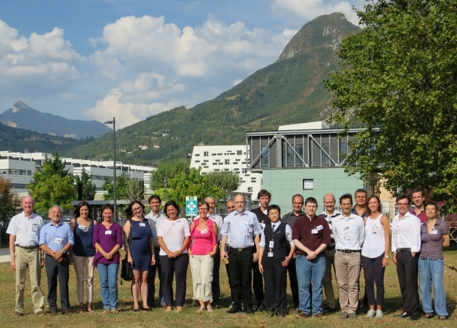
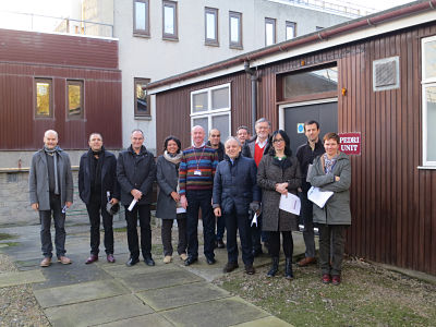
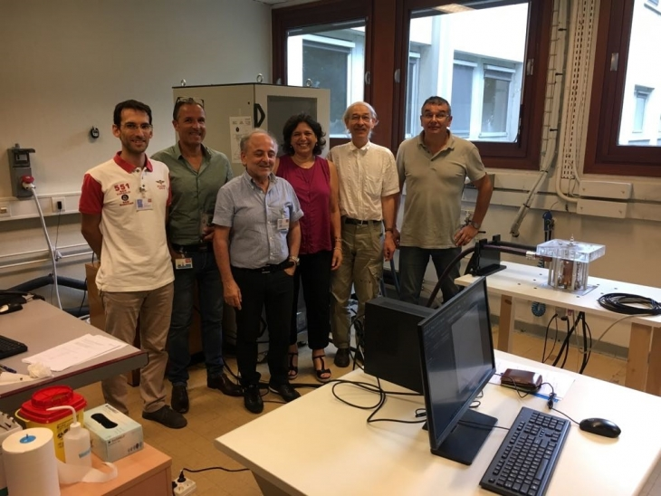
IDentIFY Group Photograph - Grenoble, September 2016
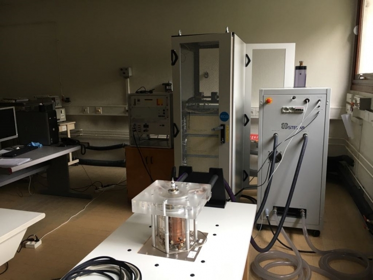
Relaxometer Installation in Grenoble July 2018
Installation of the new relaxometer in Grenoble (July 2018). Partners involved - STELAR, INSERM & CEA
IPEM John Mallard Lecture - Liverpool - June 2019
Keynote Lecture
Professor David Lurie
Institute of Phyiscs and Engineering in
Medicine (IPEM)
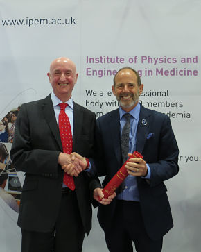
John Mallard Lecture delivered at the UK
Imaging and Oncology Congress (UKIO)
June 2019
David is presented with a certificate
by IPEM President Mark Tooley
Italian Association of Medical Physics Invited Lecture
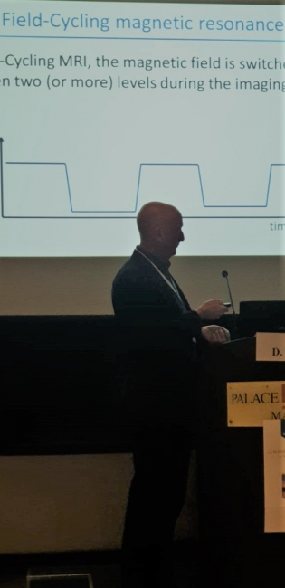
Professor David Lurie gave an invited lecture at the
meeting of the Italian Association of Medical Physics
(AIFM).
The meeting was hed in Matera, Italy from 7-8th
November 2019 and titeld “The new challenges of medical physics: a bridge between innovation and
medicine.”
The conference was held to celebrate
the International Day of Medical Physics (IDMP) on 7th November 2019
Secretary of State for Health and Social Care visits next generation MRI scanner
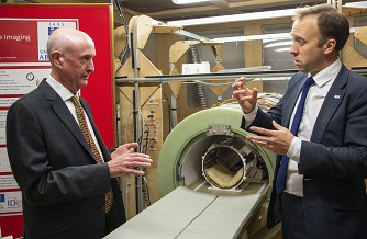
Secretary of State for Health and
Social Care, Matt Hancock, visited
the University of Aberdeen to see
first-hand the development of the
next generation of MRI scanners.
Mr Hancock visited the Medical
Physics Department where the first full body scanner was designed and
built in the 1980s
Secretary of State for Health and Social Care
Head of department, Professor David Lurie, gave Mr Hancock an
overview of the development of the next step in MRI technology,
known as Fast Field Cycling MRI.
Current MRI scanners use a large magnet along with pulses of
radiowaves to create detailed pictures of a patient’s anatomy.
Whilst current MRI machines operate on a single strength of
magnetic field, Fast Field Cycling MRI scanners are able to extract
much more information by switching the strength of the magnetic
field during the scanning procedure.
The technology has been under development for the last ten years.
Secretary of State, Local MPs, Dr Mary Joan McLeod and Professor David Lurie
The Aberdeen team behind FFC-MRI is leading a nine-strong consortium of research groups from six different countries across Europe, in a project called IDentIFY which received a €6.6 million Horizon 2020 research grant from the European Union to develop the imaging technology and bring it closer to widespread use in hospitals.
The first patients were scanned in the new machine in late 2017 and a project is currently under way to test its capabilities in detecting breast cancer.
Mr Hancock was joined by local MPs Andrew Bowie and Colin Clark.
Colin Clark MP commented: “This is a world class development and I am delighted SoS for Health was able to accept my invitation. Aberdeen Institute of Medical Science is the birth place of the MRI and this new Fast Field-Cycling MRI (FFC-MRI) scanner has national importance. MPs are always delighted to showcase the NE to Westminster ministers.”
Professor Lurie added: “It was fantastic to have the opportunity to give Mr Hancock an overview of this important technology which we believe can eventually be used in hospitals to produce more detailed information to help clinicians treat patients more effectively.”
European Research Scottish Parliament Event
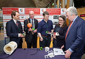
European Research Scottish Parliament Event
7th March 2019 - Holyrood, Edinburgh
From Left to Right - Ben MacPherson (Minister for Europe, Migration and International Development), Professor David Lurie, Dr James Ross, Dr Mary Joan Macleod, Lewis MacDoanld (MSP for North-East Scotland)
The IdentIFY Team and Dr Mary Joan Macleod met with the Minister for Europe at a special Scottish Parliament event to celebrate European research within Scotland.
Ben Macpherson, Minister for Europe, Migration and International Development, addressing the group, said: “Looking at the project about the future MRI development and technology, to see the collaboration of the list of countries involved, let alone the list of people, was quite remarkable and tells a story that I know all the stalls will tell as well and the whole spirit of this celebration embodies. “This event highlights the importance of research and inclusivity of research to the public and at a time where we want to be encouraging as much innovation and participation in academia and in the economy more widely, for an event to do that on such a wide scale is definitely worth celebrating again as we are this evening.”
Senior Vice-Principal of the University of Aberdeen, Professor Karl Leydecker, said: “This event is a fantastic opportunity to celebrate and see first-hand the research that the European Commission funds in Scotland as well as to mark the success of Explorathon.“The range of innovative, exciting and important projects on display tonight is testament to the importance of European-funded research and the collaboration it brings.
Explorathon takes that research and finds new ways to make it accessible and relevant to people in public places with high levels of footfall. Only through the strong links between our core universities and the dedication of all staff involved is this possible.”
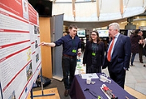
Public Engagement
British Science Week Scanners Tour 2018
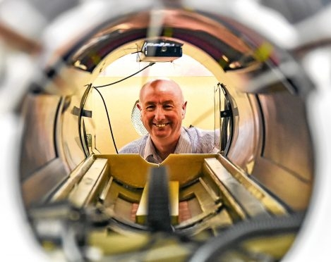
Venue: Aberdeen Royal Infirmary Art Space and Aberdeen University’s PEDRI Unit.
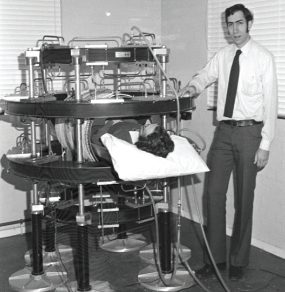
The world’s first MRI scan of a patient was done in 1980, using Aberdeen University’s “Mark-I” scanner. Now a team of scientists at the University’s Medical School has built a new kind of scanner called “Fast-Field-Cycling” MRI, which could provide earlier diagnosis of many diseases. This event is a unique opportunity to see for yourself the original Mark-I scanner in Aberdeen Royal Infirmary and to visit the laboratory where the world’s only prototype FFC-MRI scanner has been built.
Experts will be on hand to explain the scanning technology, to answer questions and to guide tours to see the scanners.
The original Mark 1 scanner
British Science Week Talks 2018
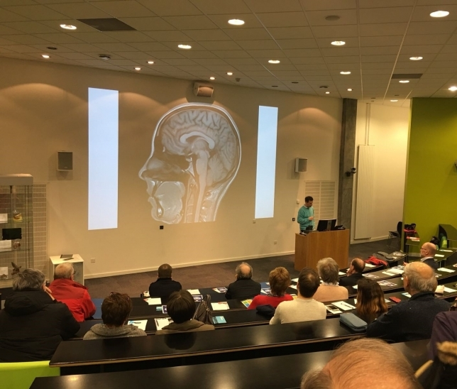
British Science week talks presented by Dr Lionel Broche (IdentIFY Project) and Dr Mary Joan MacLeod (Stroke Specialist)
What is an FFC-MRI scanner and how can it benefit patients?
Venue: Suttie Centre, Foresterhill Lecture Theatre, AB25 2ZD
Event held during the IDentIFY project half-term meetings and wokshops.
In 1980 the very first scan of a patient by MRI was carried out at the University of Aberdeen. Now a new generation of scanners has been designed and built there. Called “Fast Field-Cycling MRI”, the devices switch rapidly between strong and weak magnetic fields, generating extra diagnostic information for the doctors who read the scans. Hear from the team behind this new technology.
They will explain how the FFC-MRI scanners work, and how clinical trials using a prototype scanner are already helping medical staff to see more about their patients’ condition.
This event is led by the members of a European consortium called “IDentIFY” Improving Diagnosis by Fast Field-Cycling MRI and funded by the EU.
Cafe Sci Talk - David Lurie - 25th October 2017
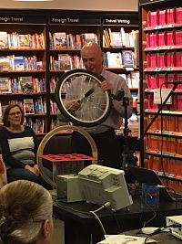
Cafe Scientifique Talk - David Lurie
Waterstones Bookshop, Aberdeen, 25th October 2017
“MRI scanning: a magnetic window to the body”:
(Note - FFC-MRI part starts at 37 minutes.)
https://www.facebook.com/CafeScientifiqueAberdeenCity/videos/1714259268646482/
Question and Answer Session:
https://www.facebook.com/CafeScientifiqueAberdeenCity/videos/1714323031973439/

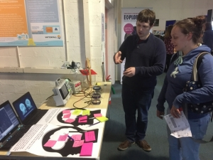
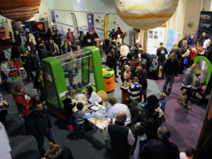
European Researchers’ Night Aberdeen - 29th September 2017
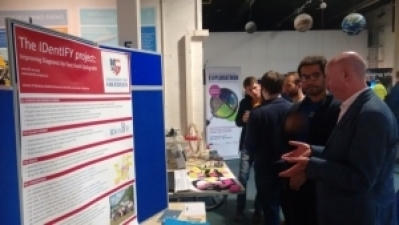
EUROPEAN RESEACHERS’ NIGHT
EXPLORATHON - ABERDEEN SCIENCE CENTRE
FRIDAY 29TH SEPTEMBER 2017
2019_ISMRM_Bilateral_Breast_coil_for_FFC_GD.pdf
Tel: 01224 552746
Email: derek.turner@abdn.ac.uk
This project has received funding from the European Union’s Horizon 2020 research and innovation programme under grant agreement No 668119
